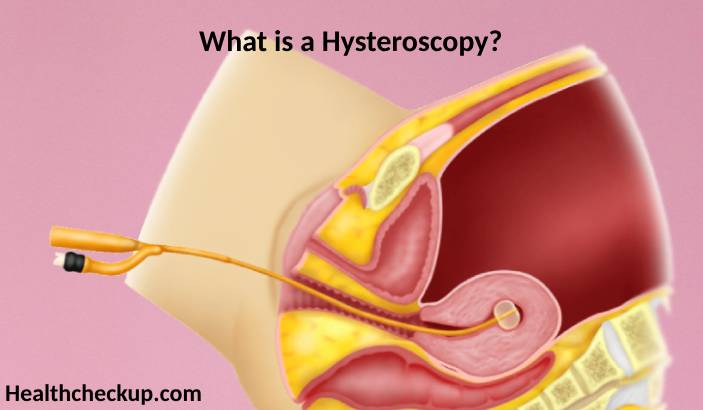
Hysteroscopy is a medical procedure that allows doctors to look inside the uterus in order to diagnose and treat causes of abnormal bleeding. The procedure is conducted using a hysteroscope, a thin, flexible, lighted tube that is inserted into the vagina to examine the cervix and inside of the uterus.
Purpose of Hysteroscopy
- Diagnostic Use: To diagnose problems in the uterus such as abnormal bleeding, fibroids, polyps, or congenital uterine anomalies which might cause infertility or repeated miscarriages.
- Surgical Use: To treat certain conditions such as removal of adhesions (scar tissue), septums, or fibroids that can occur inside the uterus.
- Postmenopausal Investigations: To investigate cases of postmenopausal bleeding to check for endometrial cancer or atrophic endometritis.
- Implantation Failure Investigation: To determine potential causes of repeated IVF failures by examining the uterine cavity.
Preparation for the Test
- Medical History Review: Disclose all medications and medical history to your doctor, especially conditions related to bleeding or use of anticoagulant medication.
- Timing: The best time for a hysteroscopy is during the first week after your period has ended, when the lining of the uterus is the thinnest.
- Pre-procedure Instructions: Depending on your specific circumstances, you may be advised to stop eating and drinking for a certain period before the procedure if anesthesia will be used.
- Post-procedure Arrangements: Since you might receive sedatives, plan for someone to drive you home after the procedure.
Procedure of Hysteroscopy
- Setting: Hysteroscopy surgery can be performed in a hospital, clinic, or a doctor’s office.
- Anesthesia: Local, regional, or general anesthesia may be used depending on the extent of the procedure and the facility.
- Insertion of Hysteroscope: The hysteroscope is inserted through your cervix into your uterus, which allows the doctor to view the uterine walls and the openings of the fallopian tubes.
- Inspection and Treatment: If any abnormal conditions are detected, instruments used through the hysteroscope can correct many of these issues during the same procedure.
Results of Hysteroscopy
- Immediate Insights: Post-procedure, your doctor can often tell you immediately about what was seen and if any immediate action was taken.
- Biopsy Results: If a biopsy was done, it may take a few days to a few weeks to get the results back from the laboratory.
- Follow-up Care: Based on the findings, further treatment or follow-up visits might be necessary to monitor your condition or the effects of the procedure.
Risks Associated with Hysteroscopy Surgery
- Common Risks: Some common, minor risks include cramping, slight vaginal bleeding, and pelvic pain.
- Infection Risk: There’s a small risk of developing an infection in the uterus or cervix.
- Uterine Perforation: Rarely, the hysteroscope can perforate the wall of the uterus, which may require further surgical repair.
- Reaction to Anesthesia: As with any procedure involving anesthesia, there is the risk of adverse reactions to the anesthetic used.
- Fluid Overload: In procedures where fluid is used to expand the uterus, there’s a risk of absorbing too much fluid, which can be serious.
Hysteroscopy surgery enables doctors to examine the uterus closely and address various uterine conditions that could affect a woman’s reproductive health and general wellbeing. It offers the benefit of being both diagnostic and therapeutic, which can eliminate the need for more invasive surgery. With its relatively low risk and high potential for providing relief and treatment, hysteroscopy is a preferred method for exploring uterine health issues, especially in the field of reproductive health and gynecology.
I specialize in writing about health, medical conditions, and healthcare, drawing extensively from scientific research. Over the course of my career, I have published widely on topics related to health, medicine, and education. My work has appeared in leading blogs and editorial columns.